Routine ankle sequences include:
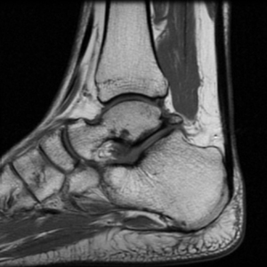
Sagittal T1
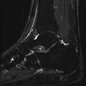
Sagittal IR
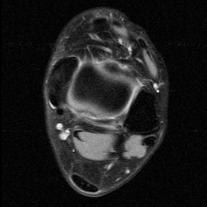
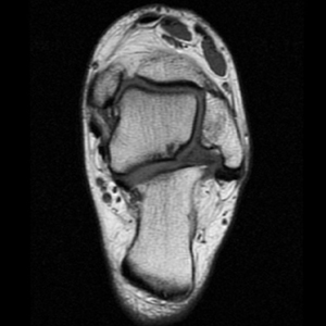
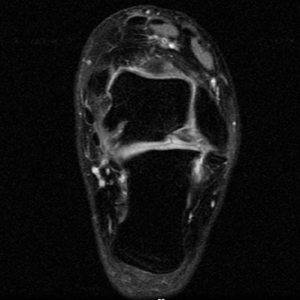
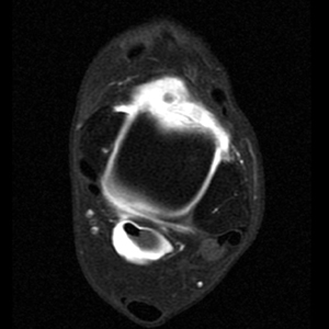
Axial PD fat suppressed FSE
Coronal Oblique T1
Coronal Oblique PD FSE fat sat or GRE
Ankle Arthrogram
Sagittal T1 and IR allow for the evaluation of the Achilles tendon, plantar aponeurosis, talar dome, subtalar facets and Sinus Tarsi.
Axial PD fat suppression evaluates the tendons and ligaments of the ankle particularly after acute/subacute injuries. It also is sensitive to talar dome osteochondral defects. Alternatively, a T2 sequence can be used to eliminate magic angle artifact that may occur as the tendons travel around the malleolar turns.
The coronal oblique T1 sequence follows the tendons of the ankle around the malleolar turns and also evaluates the medial ankle ligaments.
The coronal oblique PD fat suppressed sequence follows the tendons of the ankle around the malleolar turns and is particularly important in evaluation of the Posterior tibialis tendon.
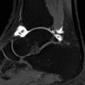
Ankle MR Arthrogram
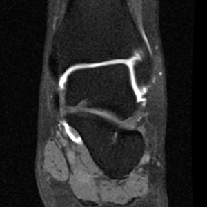
Arthrogram sequences of the ankle evaluate the talar cartilage and allow for staging of ostechondral defects of the talar dome. They can also better evaluate the joint capsule and its boundaries such as the anterior talofibular ligament.