Knee Sequences
.jpg)
Sagittal PD FSE
Sagittal PD fat suppressed CSE
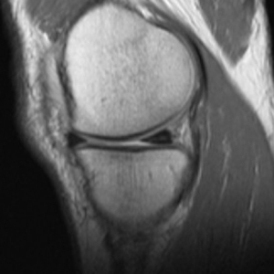
Coronal PD fat suppressed FSE
Axial IR or 3T PD fat sat FSE
Additional evaluation may include arthtographic sequences:
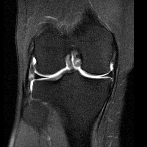
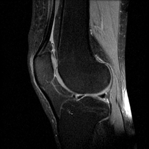
Sagittal Proton density FSE:
- The Sagittal PD FSE sequence is designed to image the Anterior Cruciate Ligament (ACL). The orientation is a sagittal oblique along the orientation of the ACL.
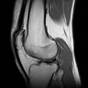
- Other structures that are well seen include the Posterior Cruciate ligament (PCL) and the extensor mechanism tendons
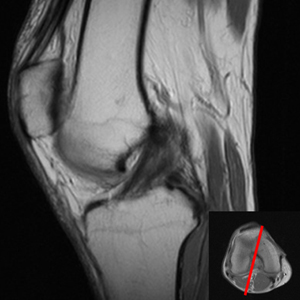
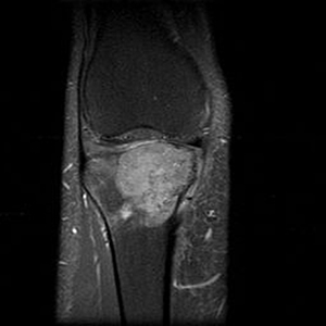
Sagittal PD fat suppressed CSE
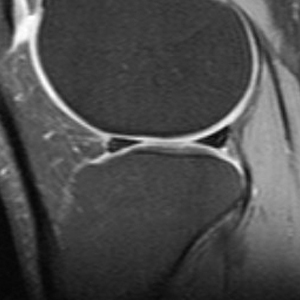
- The Sagittal PD fat suppressed CSE sequence is designed to image the menisci. Cartilage is also well visualized with this sequence, obviating the need for dedicated gradient sequences on routine studies.
- FSE techniques can introduce blur. Though conventional spin echo (CSE) sequences take longer to acquire, they are the most accurate for meniscal pathology. (rollover for PD FSE vs PD CSE fat sat)
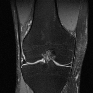
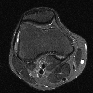
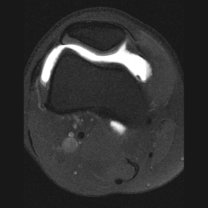
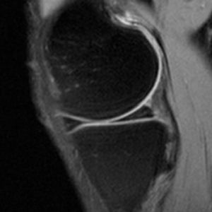
Coronal and Axial sequences

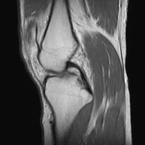
- The Coronal PD fat suppressed sequence is designed for the evaluation of the collateral supporting structures, especially when an injury is acute/subacute. Meniscal tears are also often detectable in the coronal plane.
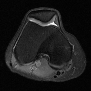
- Inversion recovery (above left) is the most sensitive sequence for detection of bone marrow edema and in the axial plane also allows for evaluation of the patellar cartilage.
- 3.0 T PD fat saturation (above right) allows for more cartilage detail.
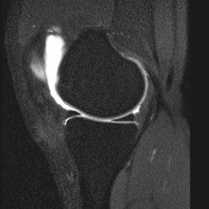
Knee Arthrogram
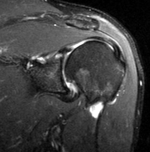
- The T1 fat suppressed post arthrogram sequence is most beneficial for evaluation of post-operative menisci. Intra-articular gadolinium helps differentiate between post-operative high signal that can be seen on PD sequences versus a retear of the meniscus. The gadolinium will enter a retear in the meniscus. The sequence can also help in evaluation of cartilage and ACL reconstructions.