Since hip pathology is often bilateral (e.g., osteoarthrosis, avascular necrosis), the routine hip protocol uses a large field of view (torso or body coil). In cases where pathology is unilateral (such as a labral tear during an MR arthrogram) a smaller field of view should be used by employing a dedicated surface coil to keep SNR acceptable while increasing spatial resolution.
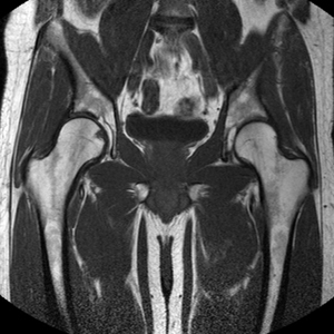
Coronal T1 both hips
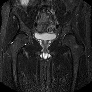
Coronal IR both hips
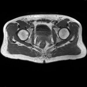
Axial T1 both hips
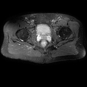
Axial PD fat suppressed both hips
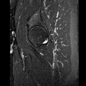
Sagittal PD fat sat affected hip
Additional Sequence:
Hip Arthrogram
Coronal Hip Imaging
Coronal T1 large field of view allows for evaluation of both hips simultaneously even though the patient may be symptomatic in only one hip.
Coronal IR large field of view allows for detection of abnormal fluid in both hips which may be seen in avascular necrosis, stress fractures, muscle tears or perilabral cysts.
Axial Hip Imaging:
Axial T1 large field of view allows for evaluation of both hips simultaneously, particularly the acetabulae.
Axial PD fat suppressed is sensitive to fluid that may be present with avascular necrosis or stress fractures, while maintaining high a SNR.
Sagital Hip Imaging:
Sagittal PD fat suppression of the affected hip is sensitive to fluid that may be present with avascular necrosis or stress fractures. Notice the decreased SNR. This is due to a combination of a large coil and decreased field of view. A large coil was used to image both hips initially in the coronal and axial planes and was not changed midstream through the exam.
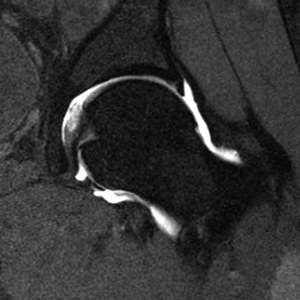
Hip MR Arthrogram:
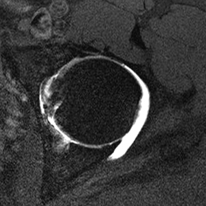
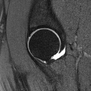
For dedicated imaging of a single hip during arthrogram, a smaller field of view with a dedicated coil or flexible surface coil is used, allowing for visualization of smaller structures such as the labrum. Notice the decreased SNR due to the smaller field of view.