Wrist imaging requires a small dedicated surface coil that allows for evaluation of very small structures such as the triangular fibrocartilage and scapho-lunate interval ligament.
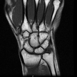
Coronal T1
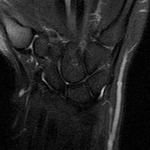
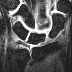
Coronal IR
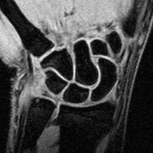
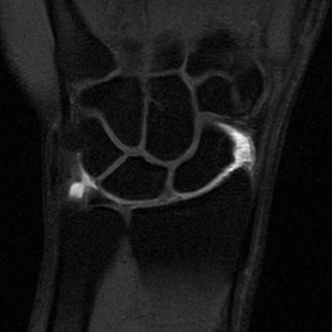
Coronal gradient
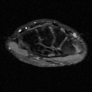
Axial PD fat suppression
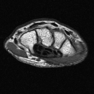
Axial T1
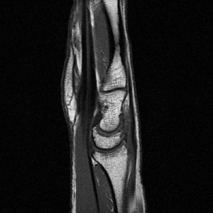
Sagittal T1
Additional Sequence:
Microscopy Coil
Wrist Arthrogram
Coronal imaging
Using a dedicated surface coil and small field of view, coronal T1 imaging evaluates bone marrow signal (e.g., increased in avascular necrosis) and the relationship of the osseous structures to each other (e.g., scapholunate disassociation).
Inversion Recovery is sensitive to pathological fluid as may be seen in an occult scaphoid fracture.
Coronal spoiled 3D gradient fat suppressed sequences are capable of very thin slice thickness which are especially well suited to imaging the small structures of the wrist. Gradient sequences also evaluate the cartilage.
Use of a 25mm microscopy surface coil allows for even thinner gradient slice thickness for dedicated evaluation of the scapholunate interval ligament and the triangular fibrocartilage complex.
Axial and Sagital imaging:
Axial T1 evaluates the tendons of the wrist and carpal tunnel, including the flexor retinaculum.
Axial PD fat suppressed evaluates the tendons of the wrist and carpal tunnel, including the median nerve.
Sagittal T1 evaluates the tendons, bone marrow and relationships between the osseous structures.
Wrist MR arthrogram:
With a radio-carpal compartment injection, post intra-articular T1 fat suppressed sequences evaluate the integrity of the interosseus ligaments and triangular fibrocartilage.