Elbow imaging requires protocols and coil selection that can accommodate both large (triceps/biceps tendons) and very small structures (collateral ligaments).
Sagittal T1
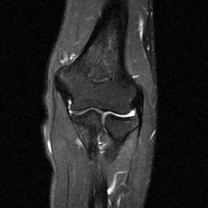
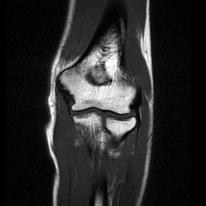
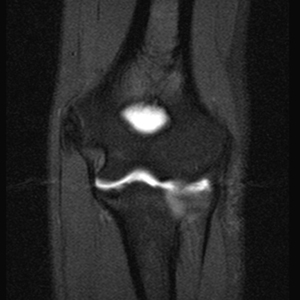
Coronal PD fat suppressed
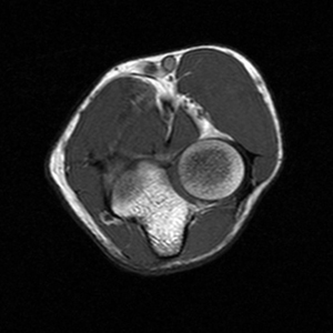
Axial T1
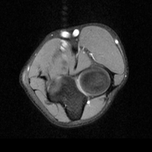
Axial PD fat suppresse
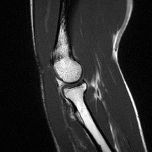
Sagittal T1
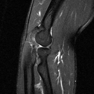
Sagittal PD fat sat
Additional Sequence:
Elbow Arthrogram
Coronal imaging
Coronal T1 and PD fat suppressed sequence are well suited for evaluation of collateral ligament and common extensor/flexor tendon group patholgy as well as epicondylitis.
Axial imaging
Axial T1 and PD FSE fat suppressed sequences evaluate the tendons of the Biceps Brachii and Brachiallis muscles transversely as they insert onto the Radius and Ulna respectively. The distal Triceps tendon is also well evlauated in this plane.
Sagital imaging
Sagittal T1 and PD FSE fat sat sequences evaluate the tendons of the Biceps Brachii and Brachiallis muscles as they travel distally to instert onto the Radius and Ulna respectively. They also help evaluate the Radial head for radiographically occult fractures. The distal Triceps tendon is also well evlauated in this plane.
Elbow MRI arthrogram
Coronal T1 fat saturated arthrogram is useful for evaluation of the collateral ligaments and cartilage surfaces.